This page presents educational information originally published by the Tennessee Breast Center. It is general knowledge, not medical advice — read our full disclaimer.
Breast Tenderness (Cyclic Mastalgia)
The breast tissue cycles hormonally during the menstrual phase, similarly to the uterus. The breasts increase in size and volume by 50% approximately two weeks prior to menses, often heralded by breast tenderness (mastalgia). The breasts usually return to their premenstrual state approximately one week after menses. This is often called mastitis or fibrocystic disease — actually a condition, since 90% of women have fibrocystic changes — and represents a normal state of the breast.
The symptoms can often be alleviated by reducing caffeine intake (coffee, tea, cola and chocolate), taking vitamin E, or taking Evening Primrose Oil, generally within three to four months. Ninety percent of the time, breast tenderness is benign (not malignant). Breast cancer is usually painless.
Breast Tenderness (Non-cyclic Mastalgia)
Generally seen in older women and not related to the menstrual cycle. Non-cyclic mastalgia may be related to a variety of medications (estrogen replacement, lanoxin, pepcid, thiazide diuretics, and so on) or to arthritic changes of the thoracic spine. Improvement occurs with anti-inflammatories.
Fibroadenoma
These benign tumors usually present as a mass in the breast during adolescence and young adulthood. The masses can enlarge and become tender during the menstrual cycle and can become quite large. With increasing age, fibroadenomas may calcify and, with this calcification, take on a typical mammographic appearance. Rarely are fibroadenomas associated with malignancy.
Lactating Adenomas
Appearing during pregnancy or lactation, they likewise present as a mass (lump) in the breast and are similar to fibroadenomas.
Nipple Discharge
Normally, ten to fifteen ducts open on the surface of the nipple, and 70% of women will periodically have nipple discharge. If the discharge is cloudy green, cloudy yellow or milky, from multiple ducts in both breasts, it is usually benign. Galactorrhea (milky discharge) may be associated with hypothyroidism (low thyroid hormone) or pituitary adenomas (benign growths of the pituitary). Measuring TSH (thyroid stimulating hormone) and prolactin levels (blood tests) may identify these as potential sources. Many medications can likewise be associated with nipple discharge. Benign multiduct nipple discharge is not an indication for surgical biopsy.
Bloody or serous (clear yellow) discharge from a single duct, noted spontaneously in nightgowns or bras, is concerning for an underlying problem and should be evaluated by a physician. Only a third of these discharges are malignant, but they represent one of the warning signs of cancer. Galactography or mammary duct excision (a surgical procedure to remove the duct) generally reveals an intraductal papilloma, a benign growth of the breast ducts.
Fibrocystic Changes
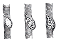
This term is generally used by clinicians to describe the induration (swelling) and tenderness associated with the menstrual cycle. Under the microscope, these changes are characterized by cysts (fluid-filled sacs) of various sizes, stromal fibrosis (scarring of the tissue surrounding the functional breast units) and a variety of proliferative lesions (increased layers of cells lining the ducts). These changes are present in 90% of women and therefore cannot be classified as a disease — they are the normal changes of the breast with increasing age.
Classification of Fibrocystic Changes
a. Nonproliferative lesions
Cysts, apocrine change, calcifications, fibroadenomas and hyperplasia (an increased number of cell layers lining the breast ducts — more than two but no more than four layers of epithelial cells). The risk of developing breast cancer is not increased in this group.
b. Proliferative lesions without atypia
Florid hyperplasia (more than four layers of epithelial cells), sclerosing adenosis and intraductal papillomatosis. Women with these changes on a breast biopsy are at slightly increased risk of developing breast cancer — 1.5 to 2 times the risk of the general population.


c. Atypical hyperplasia
Multiple cellular layers that may involve lobular or ductal elements. Women with these changes are at increased risk for developing breast cancer — 4.5 to 5 times the risk of the general population. With the addition of a positive family history of breast cancer and these findings on a breast biopsy, the risk doubles to 8 to 10 times that of the normal population.
d. Lobular carcinoma in situ (LCIS)
Although this sounds like cancer, it is still benign proliferative breast disease (hyperplasia, atypical hyperplasia). These women are at increased risk for the development of breast cancer — 15%–35% over the next fifteen years. Although once considered an indication for prophylactic mastectomy, and some women have undergone bilateral mastectomy for this condition, close clinical follow-up may suffice. Infiltrating ductal carcinoma is the most common cell type in those individuals who proceed on to breast cancer, and the tumors develop equally in either breast. Consideration should be given to chemoprevention with tamoxifen.
Fat Necrosis
Generally associated with a history of trauma, fat necrosis presents as a firm mass in the breast. Clinically and by mammography it may be indistinguishable from carcinoma of the breast. The diagnosis is usually made by breast biopsy.
Subareolar Abscess
A subareolar abscess presents with pain and redness of the nipple-areolar complex. The abscess is generally polymicrobial (more than one bacterium) and is usually seen in smokers. Treatment is by incision and drainage.
Lactational Mastitis
Seen within the first weeks after the onset of breast-feeding, the breast becomes inflamed (red), warm and tender in a wedge-shaped distribution. Cracked nipples usually cause this condition. The organism responsible is usually Staphylococcus aureus, and the condition responds to antibiotics. If not treated promptly, it may lead to a breast abscess requiring surgical drainage.