This page presents educational information originally published by the Tennessee Breast Center. It is general knowledge, not medical advice — read our full disclaimer.
How a Breast Mass Is Evaluated
The evaluation of a breast mass at a breast center may differ from a general physician’s office. If a patient has had a mammogram, the films are requested and reviewed. If films are not available, a mammogram may be requested based on the patient’s age — a younger patient may undergo sonography (ultrasound) first, to characterize the lesion.
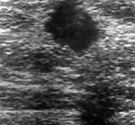
Are there any abnormalities on the mammogram? Sonography may be performed to determine whether the mass is cystic or solid. If the mass is a simple cyst, the evaluation is complete — it is a benign process (not cancer). If the cyst is tender or quite large, it can be aspirated with relief of those symptoms. Half of aspirated cysts recur; if air is injected into the cyst after aspiration, only 2% recur.
When a Cyst Needs More Attention
If the fluid retrieved is bloody, or the cyst incompletely resolves, injecting air into the cyst and repeating the mammogram may reveal an intracystic lesion (a growth inside the cyst). A bloody aspirate or an intracystic lesion indicates the need for a breast biopsy.
If the Mass Is Solid: Types of Breast Biopsy
If the mass is solid on sonography, or suspicious mammographically, your physician may recommend a breast biopsy. There are several types:
| Type | How it works |
|---|---|
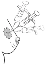
| Fine needle aspiration biopsy (FNAB) | A small needle is inserted into the mass and passed in several directions to collect cells for the pathologist. A malignant result is 100% accurate; a benign result is only 64% accurate, so close follow-up is warranted. Accuracy depends on the skill and adequacy of the sampling and the experience of the cytopathologist. |
| Open surgical biopsy | Usually an outpatient procedure under local anesthesia. If the concern is a mammographic lesion, localization (hook-wire or needle localization, guided by mammography or sonography) occurs before the biopsy. Usually results in complete removal of the lesion. |
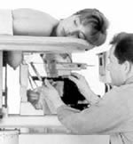
| Stereotactic core biopsy | A computer-guided core needle biopsy that samples the lesion using mammographic guidance. |
| Sonography-guided core biopsy | A core needle biopsy guided in real time by ultrasound. |
The Pathology Report
The pathology report takes several days to return. Remember: 80%–90% of lesions detected by clinical examination or mammography are benign.
Related: early detection and screening guidelines and the step-by-step breast self-exam guide.