This page presents educational information originally published by the Tennessee Breast Center. It is general knowledge, not medical advice — read our full disclaimer.
Historical Aspects of Surgical Management
We have known about breast cancer for thousands of years. The Smith Papyrus discusses breast cancer in 3000 BC; it was treated with a variety of topical agents and poultices, and understandably those women did not survive. There were early attempts at surgical treatment in the early 1100s.

Anesthesia was not developed until 1846, and antibiotics not until 1927. The breast was rapidly amputated with women awake; if the operation itself was not fatal, about 20% of these women died from infection. In 1894, William Halsted, M.D. (the father of American surgery) developed the radical mastectomy, removing the breast, the chest wall muscles and the lymph nodes under the arm. It was a severely deforming operation and women had lasting disability involving the arm, but it produced the first long-term breast cancer survivors. Women typically presented with large tumors or extensive disease — mammography was not utilized until the 1970s. This remained the standard operation through the late 1960s.
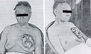
In the 1970s, treatment moved to the modified radical mastectomy — removing the breast while preserving the chest wall muscles and sampling the axillary nodes. These women survived just as long as those who had radical mastectomies, and the modified radical mastectomy became the standard operation, remaining an option today.

Italian investigators began conservative breast surgery (lumpectomy and irradiation) in the early 1970s, initially selecting women whose mass was under 2 cm with clinically normal lymph nodes. A quarter of the breast was removed, the lymph nodes sampled and the breast irradiated — and these women survived just as long as those treated with modified radical or radical mastectomy. In the United States, the selection criteria grew to masses under 4 cm, with clinically enlarged lymph nodes still acceptable. The whole breast must be treated in some fashion — either removed or irradiated — because breast cancer can be multifocal (involving multiple areas of the breast 23%–55% of the time).
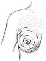
Not every woman is a candidate for breast conservation. The following may exclude some women:
- Certain cell types of breast cancer are prone to local recurrence when treated with conservative surgery.
- Mammographic or clinical evidence of multifocal disease (tumor involving multiple areas of the breast).
- A large tumor in a small breast — the cosmetic result of conservative surgery may be unacceptable.
- Inability to obtain microscopically free margins around the tumor.
- Relative contraindications include collagen vascular disease and pregnancy.
Surgical Options Currently Recommended for Breast Cancer
- Simple mastectomy or lumpectomy (with irradiation) without axillary dissection — for in-situ lesions. Fewer than 1% of patients with intraductal lesions have positive lymph nodes, so the nodes are not sampled.
- Conservative breast surgery — lumpectomy, axillary dissection and irradiation.
- Modified radical mastectomy — removal of the breast and axillary dissection (sampling the lymph nodes).
- Modified radical mastectomy with primary reconstruction — removal of the breast, axillary dissection and rebuilding the breast at the time of mastectomy. Reconstruction can also be accomplished at any time after mastectomy.
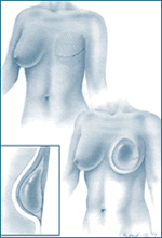
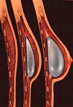
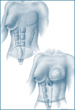
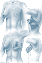
Stage I and stage II lesions are treated in the fashion above. Stage III lesions are generally treated with chemotherapy first, then surgery and possibly irradiation. Tumors with more than four positive lymph nodes generally receive adjuvant irradiation in addition to surgery and chemotherapy.
Sentinel Node Biopsy
Sentinel node biopsy is a technique that uses a radioactive tracer or blue dye injected into the tumor. The contrast material is absorbed by the lymphatic system and travels to the first lymph node draining the tumor — the sentinel node. If the sentinel node is negative, a woman may not need a more extensive axillary dissection, avoiding potential complications such as lymphedema (swelling of the arm secondary to lymphatic obstruction).
Interactive demo: the original 2002 site included a Flash animation of a sentinel node biopsy. You can still run it below in the browser, on demand.
Chemotherapy
Prior to 1988, if a woman had pathologically negative axillary nodes (no tumor in the lymph nodes under the microscope), chemotherapy was not recommended. In 1988 the NIH recommended that all women receive chemotherapy. Since 70% of women achieve long-term disease-free survival with surgery alone, that recommendation meant 100% of women would receive chemotherapy when only 30% would benefit. It was based on the observation that some women with stage I disease recur 15–20 years later with metastatic disease — suggesting breast cancer may be a systemic disease (microscopic disease elsewhere) at the time of diagnosis.
Nationally, the recommendation has since been modified: chemotherapy for node-negative women is recommended based on prognostic indicators. If the tumor is larger than 1 cm and the prognostic indicators suggest an increased recurrence rate, chemotherapy is recommended. Essentially all patients with positive axillary nodes receive chemotherapy.
A variety of agents are used, including cyclophosphamide, methotrexate and 5-fluorouracil (CMF), Adriamycin (doxorubicin) and cyclophosphamide (AC), paclitaxel (Taxol), docetaxel (Taxotere) and tamoxifen. The medical oncologist selects these based on the prognostic indicators, the patient’s age and underlying medical condition. Chemotherapy generally begins within two weeks of surgery and continues for several months. It can reduce the risk of recurrence by 30%–40%, and reduce the risk of cancer in the other breast by a similar percentage. Side effects vary with the agents used and the individual.
Irradiation
Irradiation is used to treat the breast after conservative surgery, for consolidation of extensive local disease (large tumors or more than four positive axillary nodes) or for local recurrence. It destroys the microscopic residual disease that remains after surgery or is not clinically apparent. Like surgery it is a local treatment, in contrast to chemotherapy, which is systemic. Side effects include erythema of the skin (sunburn-like), progressing to brawny induration (thickening and increased pigmentation), some shrinkage of breast tissue after conservative surgery, and fatigue in some women. Irradiation generally follows chemotherapy when both are given. It can also be used for palliation (relief of symptoms such as bone pain) with recurrent disease.
Local Recurrence and Systemic or Metastatic Disease
At the time of diagnosis, breast cancer may already be a systemic disease — there may be microscopic or macroscopic (clinically detectable) disease elsewhere. This is the basis for chemotherapy recommendations. Before surgery, a chest x-ray and blood tests (liver function and serum calcium) are obtained; if these are negative, fewer than 4% of women have demonstrable disease elsewhere. A CT scan of the chest and upper abdomen and a bone scan are generally obtained during treatment as baseline studies for comparison. Routine annual imaging other than mammography is not performed, because there is no survival advantage in detecting an asymptomatic recurrence. Systemic recurrence is treated with chemotherapy.
Local recurrence after conservative surgery is treated with salvage mastectomy. Local recurrence after mastectomy is treated with wide surgical excision when possible, plus irradiation. Both are usually given additional chemotherapy.
Medical links
The following organizations publish authoritative breast health information. These links are provided for convenience and further information; they are not endorsements.