This page presents educational information originally published by the Tennessee Breast Center. It is general knowledge, not medical advice — read our full disclaimer.
The Three-Part Early Detection Routine
- Monthly breast self-examination
- Yearly physician examination
- Mammography according to the American Cancer Society
The American Cancer Society guidelines for the detection of breast cancer in asymptomatic women (as published by the center) are:
| Age | Recommendation |
|---|---|
| 20 and older | Perform breast self-examination every month. |
| 20–39 | Physical examination of the breast every three years by a health care professional. |
| 40 and older | Physical examination of the breast every year by a physician, physician assistant, nurse or nurse practitioner. |
| 40 and older | Mammogram every year. |
Monthly Breast Self-Examination
Breast cancer feels like a hard rock in the breast 78% of the time; the other 22% of the time it feels like rubbery thickening. Women find 80%–90% of the masses in their own breasts. Women who routinely perform breast self-examination can detect a mass at one-half inch in size, while physicians typically detect tumors at one inch. Self-examination is effective in detecting 70% of breast abnormalities — it makes you your own breast expert.
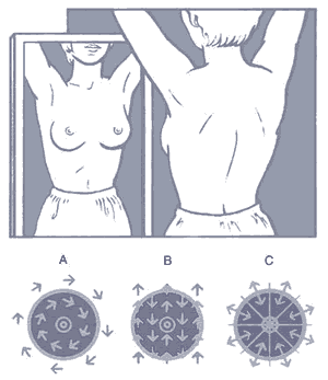
The best time for self-examination is a week after your period; if you do not have periods, pick a day of the month. The exam has three parts:
- Visual inspection
- Examination upright
- Examination lying down
For the complete walk-through with illustrations, see our step-by-step breast self-exam guide.
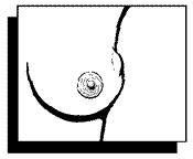
Nipple changes
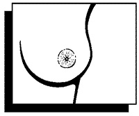
The nipples should point in the same direction. As women age, nipples tend to move down and out. Look for retraction of a nipple, or a change on one side compared with the other, plus scaling or redness of the nipple-areolar complex. Check for discharge: bloody or clear-yellow discharge from a single duct in one breast needs physician evaluation, as a third of such discharges may represent an underlying malignancy. Multiple-duct discharge involving both breasts that is cloudy green, yellow or milky is usually benign.
Skin changes
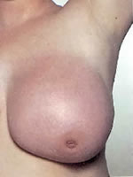
Notice the color and consistency of the skin. Redness, thickening, or prominence of the hair follicles (peau d’orange) needs evaluation by a physician.
Breast contour changes
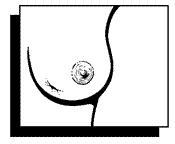
Looking at the breasts, watch for abnormal bulges or skin dimpling; these can be more prominent with the arms extended above the head.
Examination upright
Several examination patterns exist; the pattern matters less than examining consistently. Generally recommended in the shower or tub with soap, the breast is examined systematically with the flat of the fingertips, using firm pressure to feel the internal structures. Examine the right breast with the left hand and vice versa; soap or lotion makes this easier. The breast tissue is arranged in lobules — groups like the petals of a flower. If you feel these separations, examine in a radial or starburst pattern, and compare one breast to the other. Any mass or thickening not present in the opposite breast should be reported to your physician or breast center.
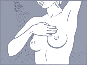
Check the lymph nodes in your armpit (axilla) — small firm kernels, like those that develop in your neck with a sore throat. If you notice any masses, see your physician: 10% of breast cancer may present as an axillary node alone.
Examination lying down
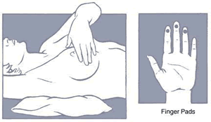
Repeat the same examination lying down, when the breast tissue is compressed against the chest wall, allowing you to feel the internal structures. Many masses can only be felt in one position — that is why both upright and lying-down examinations are recommended.
Physician Examination
If you notice a change during self-examination, or as part of your yearly well-woman exam, your physician should perform a breast examination. Physician evaluation detects breast abnormalities about 70% of the time. Your evaluation should include:
- Breast history
- Assessment of risk factors
- Breast examination — visual inspection; upright examination; examination lying down; and assessment of the axillary and supraclavicular nodes (above the collar bone)
Mammography
Mammography is a radiological technique used to detect breast abnormalities in symptomatic and asymptomatic patients. The first dedicated mammogram units were developed in 1969 and clinical trials began in the early 1970s. Compression stabilizes the breast and spreads its internal structures, allowing a lower radiation dose for imaging. The dose from a mammogram is 0.1 rad per view — the risk is negligible (you would have to live to age 7,500 with a yearly mammogram for a one-in-a-million chance of developing breast cancer from mammography).
Mammography can reduce the risk of dying from breast cancer by 30%–40%, and can detect breast cancer about three years sooner than a physical examination. The key to survival is early detection. There are two types:
- Screening mammogram — two views of each breast in asymptomatic women, intended to detect cancer before it becomes clinically apparent.
- Diagnostic mammography — additional views obtained after a screening mammogram shows an abnormality, or for patients with known breast conditions such as a mass, implants or a history of breast cancer.
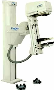
The American Cancer Society recommends that all women over forty have a yearly mammogram. Women at increased risk from a premenopausal family history may need to begin screening earlier. Women treated with conservative breast surgery need diagnostic imaging of the affected breast every six months for the first two years, then yearly diagnostic imaging. All breast facilities are required to be ACR (American College of Radiology) and FDA accredited.
Next step: learn the self-exam routine step by step.